
The electrocardiogram, usually called an ECG, is one of the best-known tests used to evaluate the heart. It is simple, quick and non-invasive: it records the heart’s electrical activity and turns it into a trace that a physician can interpret. This is why it is used for symptoms, prevention and health surveillance at work.
Each heartbeat is regulated by electrical impulses that coordinate the different parts of the heart. Small electrodes on the skin—usually on the chest, arms and legs—record these impulses and provide information about rhythm, rate and possible changes in electrical conduction.
What is an ECG?
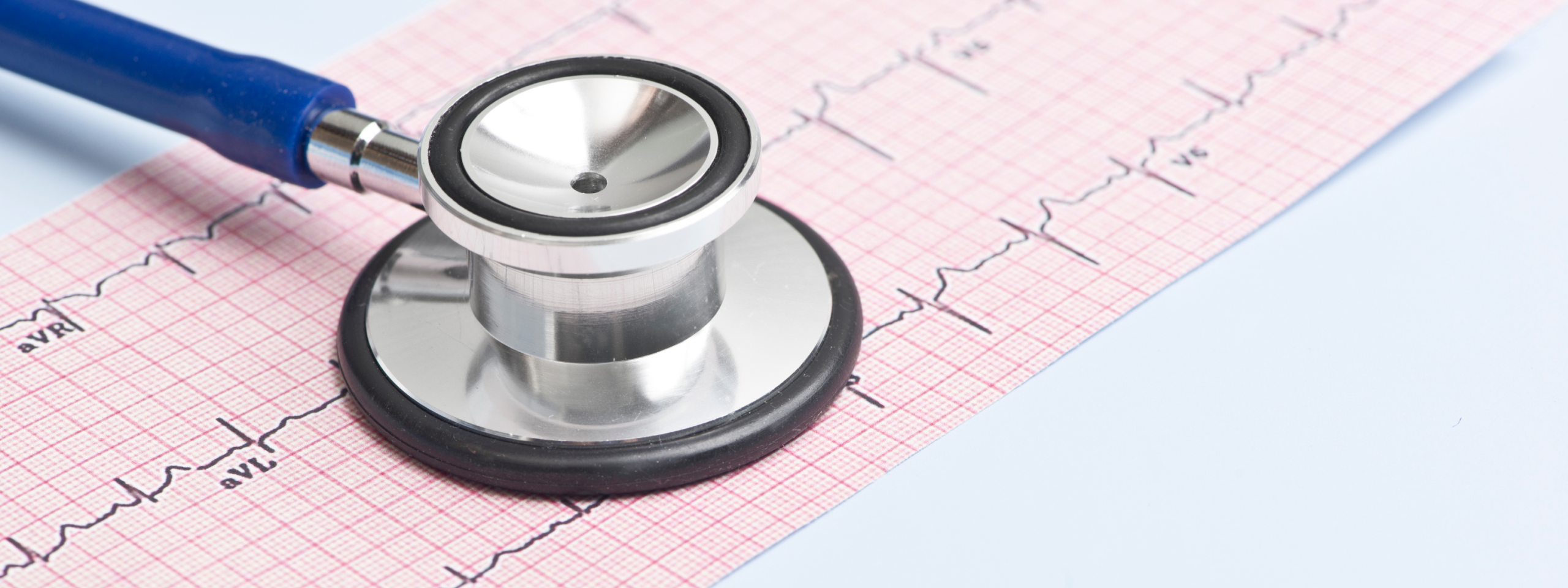
An ECG is a diagnostic test that measures and records the heart’s electrical activity. The patient usually lies down and relaxes while electrodes collect the signals produced by each heartbeat. The signals appear on paper or a screen as an electrocardiographic trace.
An ECG can show rhythm abnormalities, signs compatible with cardiac strain, conduction changes or the effects of some medicines. If there is a clinical concern, it may need to be combined with an echocardiogram, stress test, Holter monitoring or specialist assessment.
What is an ECG used for?
One common use is evaluating heart rhythm: a physician can see whether the heart beats regularly, whether the rate is high or low and whether arrhythmias are present.
It may also be requested for palpitations, chest pain, dizziness, fainting, unusual fatigue or breathing difficulty. The trace can help determine whether symptoms may be cardiac or whether further tests are needed.
The ECG is also useful for monitoring people with a known cardiac condition or taking medicines that affect rhythm, and before surgery, medical procedures or particularly demanding physical activity when a preliminary assessment is appropriate.
How the test is performed
For a resting ECG, the patient uncovers the chest, wrists and ankles or the areas needed for electrode placement. The patient lies on the examination table while small adhesive sensors are connected to the electrocardiograph. It is important to remain still and breathe normally.
The recording itself usually takes a few seconds, although preparation and saving the trace take a few minutes. The electrodes are then removed and normal activities can usually resume.
No complex preparation is normally needed and fasting is not usually required. The physician should know about medicines, known conditions and symptoms, including therapies that may affect heart rhythm.
Types of electrocardiogram
The resting ECG is the standard test, performed while the person is still and relaxed.
A stress ECG records cardiac activity during controlled exercise on a treadmill or exercise bike. It may be useful when symptoms appear during exertion.
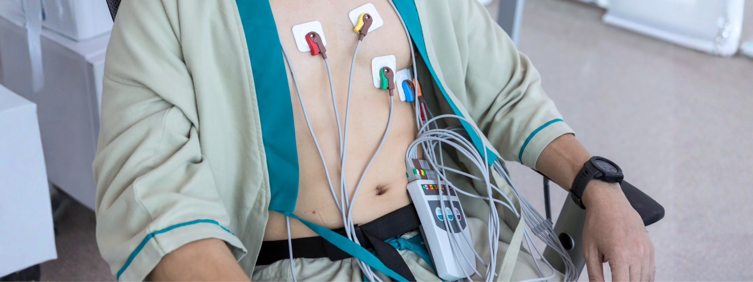
A Holter ECG is a longer recording, often 24 or 48 hours or more. The patient wears a small portable device while living normally, which is useful when symptoms are intermittent.
Each type has a specific purpose. The choice depends on symptoms, risk factors and the physician’s assessment.
What can an ECG show?
An ECG can show whether the rhythm is regular, whether the rate fits the person’s condition and whether there are conduction changes or signs that need further assessment.
It may identify patterns compatible with arrhythmias, conduction disorders, cardiac muscle strain, previous cardiac events or electrolyte imbalance. It can also help assess the effect of medicines that change specific intervals.
The trace must always be read in the context of the person’s symptoms, history, age, therapies and risk factors. A normal trace does not exclude every heart problem, and an abnormality does not automatically mean a serious disease.
ECG and cardiovascular prevention
The ECG can support prevention when included in a broader health assessment. Blood pressure, cholesterol, blood glucose, weight, smoking, inactivity, stress, sleep and family history all contribute to cardiovascular risk.
In this context, the ECG can be a useful tool for identifying signals that deserve attention.
ECG and occupational health
The ECG also has a place in occupational health when the competent physician considers it appropriate within the company health protocol. The goal is not to perform a generic cardiac diagnosis on every worker, but to assess fitness for the specific role and protect health in relation to occupational risks.
Health surveillance is defined around the risk assessment and the characteristics of the job. Correctly including tests in the protocol means real prevention rather than a formal exercise.
For workers, it is an opportunity to identify conditions that may need further assessment and to receive useful health advice.
The ECG according to Skema
The ECG is a simple, safe and widely used test that provides important information about the heart’s electrical activity. Its value concerns diagnosis and monitoring as well as prevention, especially when it is part of a complete medical assessment.
In occupational health, the ECG can be relevant within health surveillance when the competent physician considers it consistent with the role’s risks and the worker’s protection. It should not be interpreted in isolation, but together with the medical visit and other assessments.
Skema HSE takes an integrated, advisory and multidisciplinary approach to workplace health and safety. We support companies with regulatory duties and promote a continuous safety culture focused on people and better working conditions.